Blog
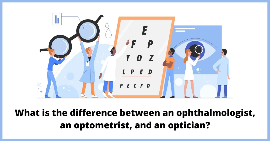
Knowing the difference between the various specialties in the eye care industry can be confusing, especially given the fact that they all start with the same letter and in many ways sound alike.
So, here’s a breakdown of the different monikers...
Read more: Ophthalmologists, Optometrists, Opticians - What's the Difference?
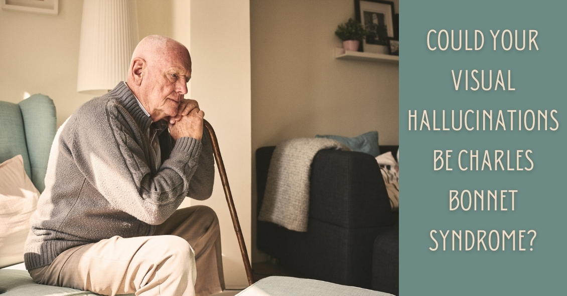
Who is Charles Bonnet? He was a Swiss naturalist, philosopher, and biologist (1720-1793) who first described the hallucinatory experiences of his 89-year-old grandfather, who was nearly blind in both eyes from cataracts. Charles Bonnet Syndrome...
Read more: Vision Hallucinations and Charles Bonnet Syndrome
