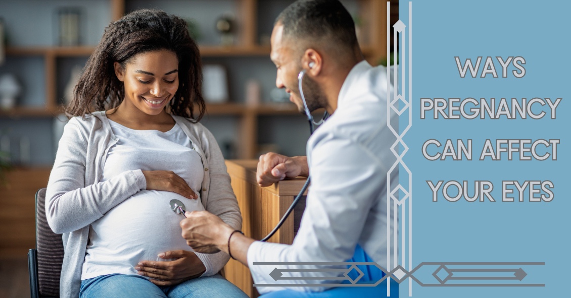
There are many opinions on the topic of texting and driving. The goal of this blog is to explore the effects on vision during texting.

So why does texting make you more likely to crash from a visual perspective? The problem lies in distraction from driving. For example, it takes a fast texter approximately 20 seconds to read and reply to a text. At 55 mph on the highway, a driver glances away from the road for approximately one-third of a mile. When the driver is focusing on their screen, this essentially gives the driver tunnel vision, causing the visual system to essentially use peripheral vision for driving. Your central vision is used to detect depth perception, detail, and colors such as red or green. So when texting, your depth perception, or 3-D vision, is altered and if cars are stopped ahead or closing in rapidly, it's not as easily detected. Colors, such as red brake lights or traffic signals, are not as easily noticed.

Next time you encounter situations with texting and driving, know that the visual system was designed to perform advanced visual perception while using central vision. This includes detail vision, depth perception, and color vision....all of which are placed on hold while texting and driving.

For more information on texting and driving see:

www.itcanwait.com

US Dept. of Transportation

www.AAA.com


The content of this blog cannot be reproduced or duplicated without the express written consent of Eye IQ.

We commonly see patients who come in saying that their eye is bleeding.

The patients are usually referring to the white part of their eye, which has turned bright red. The conjunctiva is the outermost layer of the eye and contains very fine blood vessels.  If one of these blood vessels breaks, then the blood spreads out underneath the conjunctiva. This is called a subconjunctival hemorrhage.

A subconjunctival hemorrhage doesn't cause any eye pain or affect your vision in any way. Most of the time, a subconjunctival hemorrhage is asymptomatic.  It is only noticed when looking at the mirror or when someone else notices the redness of the eye.  There should not be any discharge or crusting of your lashes.  If any of these symptoms are present, then you may have another eye condition that may need treatment.  

What causes a subconjunctival hemorrhage?  The most common cause is a spontaneous rupture of a blood vessel.  Sometimes vigorous coughing, sneezing, or bearing down can break a blood vessel.  Eye trauma and eye surgery are other causes of subconjunctival hemorrhage.  Aspirin and anticoagulant medication may make patients more susceptible to a subconjunctival hemorrhage but there is usually no need to stop these medications.  

There is no treatment needed for subconjunctival hemorrhage.  Sometimes there may be mild irritation and artificial tears can be used.  The redness usually increases in size in the first 24 hours and then will slowly get smaller and fade in color.  It often takes one to two weeks for the subconjunctival hemorrhage to be absorbed.  The larger the size of the hemorrhage then the longer it takes for it to fade.

Having a subconjunctival hemorrhage may be scary initially but it will get better in a couple of weeks without any treatment. However, redness in the eye can have other causes, and you should call your eye doctor, particularly if you have discharge from the eye.

Article contributed by Dr. Jane Pan


Dry Eye Disease affects more than 5 million people in the United States, with 3.3 million being women and most of those being age 50 or over. And as people live longer, dry eye will continue to be a growing problem.

Although treatment options for dry eyes have improved recently, one of the most effective treatments is avoidance of dry eye triggers.

For some that might mean protecting your eyes from environmental triggers. To do that experts recommend using a humidifier in your home, especially if you have forced hot-air heat; wearing sunglasses when outside to help protect your eyes from the sun and wind that may make your tears evaporate faster; or being sure to direct any fans  - such as the air vents in your car - from blowing directly on your face. For others, it may mean avoiding medications that can cause dry eyes.

There is one other trigger that may need to be avoided that doesn’t get as much notice: the potentially harmful ingredients in cosmetics.

Cosmetics do not need to prove that they are “safe and effective” like drugs do. The FDA states that cosmetics are supposed to be tested for safety but there is no requirement that companies share their safety data with the FDA. There are also no specific definition requirements for labeling cosmetics as “hypoallergenic,” “dermatologist tested,” “ophthalmologist tested,” “sensitive formula” or the like, making most of those labels more marketing than science.

Things to watch out for in your cosmetics if you have dry eye include:

Preservatives

Preservatives are important to prevent the cosmetics from becoming contaminated but many are known to exacerbate dry eye. Common preservatives in cosmetics that could be adding to your dry eye problems (Periman and O’Dell, Ophthalmology Management August 2016) are: BAK (Benzalkonium chloride); Formaldehyde-donating (yes, Formaldehyde!) preservatives (often listed as DMDM-hydantoin, quaternium-15, imidazolidinyl urea, diazolidinyl urea and 2-bromo-2-nitropropane-1,3-diol); parabens; and Phenoxyethanol.  All of these preservatives in sufficient quantities can cause ocular irritation or inhibit the function of the Meibomian Glands that produce mucous that coats your tear film and keeps it from evaporating too quickly.

Alcohol

Alcohol is used in cosmetics mostly to speed the drying time but the alcohol can also dry the surface of the eye.

Waxes

Waxes can block the opening of the Meibomian Glands along the eyelid margin. If these glands are blocked they will not be able to supply the mucous and lipids necessary to the tear film to prevent it from drying too quickly. If you have trouble with dry eye it would be advisable not to apply eye liner behind the eyelashes along the lid edge where the Meibomian gland openings are.

Anti-aging products

While these may be safe and effective for the skin of the face they should not be used around the eyes. Most of these products contain some form of Retin A. These products have been shown to be toxic to the Meibomian glands and could be contributing to your dry eyes.

These components of cosmetics do not adversely affect everyone. However, if you suffer from dry eye and are not effectively able to keep your eye comfortable and your vision clear, you should investigate your cosmetics as a potential contributor to your problem.

Article contributed by Dr. Brian Wnorowski, M.D.

The content of this blog cannot be reproduced or duplicated without the express written consent of Eye IQ

Despite requests that patients bring their current glasses to their office visit, many show up without them.

Sometimes it’s an oversight: “I was rushing to get here and forgot them”; “I left them in the car”; “I picked up my wife’s glasses instead of mine by mistake.” Doctors have heard them all.

Sometimes it is unavoidable: “I lost them”; “They were stolen”; “I ran them over with the car”; “I left them on the roof of the car and drove away and now they are gone.”

Frequently, however, it’s intentional. There is a perception by some people that if they don’t like their current glasses or feel like they are not working well for them that they are better off having their eye doctor start from scratch. “Why would I want the doctor to utilize a pair of glasses I’m not happy with as a basis or starting point for my next pair of glasses?”

But bringing your glasses to an appointment is important.

There are two main reasons for eye care professionals to know what your last pair of glasses were.

The first is to see what type of glasses they are and how you see out of them. Are they just distance? Just reading? A bifocal? A trifocal? A progressive?

Even if you feel they aren’t working for you it is essential for doctors to know the type of lens you had previously. It is also important to know how you see out of them and what the previous prescription was. This can help eye care professionals determine a new prescription that will work better for you.

The second reason doctors like to know what was in your last pair of glasses is that the majority of people who wear eyeglasses have some degree of astigmatism in their eyeglass prescription.

A significant change in either the amount or axis of the astigmatism correction from one pair of glasses to the next is often not tolerated well, especially in adults. If you make too much change from the previous prescription many people experience a pulling sensation in their eyes when they wear the new glasses. It can cause symptoms of eye strain, headaches, and distortion, making flat objects like a table look like they are slanted.

Many of the problems that occur when we try to give someone a new eyeglass prescription could be avoided if doctors knew the last prescription and how you did with it.

Anytime you are going to the eye doctor, it is essential to bring your most current pair of glasses with you to the exam--whether you love them or hate them!


Article contributed by Dr. Brian Wnorowski, M.D.

This blog provides general information and discussion about eye health and related subjects. The words and other content provided on this blog, and in any linked materials, are not intended and should not be construed as medical advice. If the reader or any other person has a medical concern, he or she should consult with an appropriately licensed physician. The content of this blog cannot be reproduced or duplicated without the express written consent of Eye IQ.

We are frequently asked if it’s wise to have cataract surgery if you have Macular Degeneration.

Let’s start with some background.

  1. Cataracts and Age-related Macular Degeneration (AMD) are both leading causes of visual impairment in the elderly population.
  2. Cataracts develop when the normal clear lens gets cloudy with age. This is correctable with cataract surgery, which involves replacing the cloudy lens with a clear, artificial lens.
  3. While cataracts affect the front part of the eye, AMD causes damage to the retina, which is the inner back lining of the eye.

In the past, there was a concern about cataract surgery causing progression of AMD. It was thought that there was an inflammatory component to AMD and the normal inflammatory response after cataract surgery may lead to AMD progression.

But studies have looked at patients who underwent cataract surgery compared to patients who didn't have cataract surgery and the progression of AMD was not significantly different between the two groups. However, those patients with AMD who underwent cataract surgery had a significant improvement in vision.

AMD patients can further be characterized as having wet or dry AMD, and only those with wet AMD require treatment. Patients with wet AMD need injections to decrease the growth of new blood vessels and reduce fluid in the retina.

A 2015 study showed that after cataract surgery, there was an increase in fluid in the retina of patients with wet AMD. Therefore, in patients with wet AMD, we usually want the wet AMD to be stabilized before the patient has cataract surgery. Sometimes an injection may be given prior to cataract surgery to prevent any inflammatory changes that may be associated with cataract surgery.

The majority of the studies on the subject conclude that it is safe to have cataract surgery even if you have AMD and in most cases there is a significant improvement in vision. Removing the cloudy lens also helps the ophthalmologist to better monitor the status of the AMD.


Article contributed by Dr. Jan Pan.

The content of this blog cannot be reproduced or duplicated without the express written consent of Eye IQ

So you’re going about your day and notice a slight twinge when you blink. It starts off as a mild awareness then proceeds to a painful feeling with every blink. You look in the mirror to see what could be causing it, and there you see a small red bump forming.

You decide to wait to see what happens and one of three things occurs. Either it gets bigger, redder, and more painful; it gets smaller and goes away; or it stays put and is no longer painful or growing in size. Let’s dive into one of the most common eye conditions we treat: hordeola (commonly known as “styes”) and chalazia.

Hordeola (or singular hordeolum), are infectious abscesses of the glands that line the eyelids. Bacteria that are naturally occurring on the eyelids and eyelashes can make their way into the gland and form what is essentially a pimple in the eyelid. If it goes untreated, hordeola can (rarely) lead to spreading of the infection throughout the eyelid (preseptal cellulitis) or even start to invade the orbit of the eye (orbital cellulitis).  At that point, infection can spread to the brain and even be life-threatening and require hospitalization. Thankfully it hardly ever gets to this point.

Treatment can consist of warm compresses, ointments, and oral antibiotics to kill the infection. Just like a pimple, it is not always necessary to be on antibiotics.  It’s actually the warm compresses that do the most good.  If you can get the mucous that is clogging the gland opening to thin out it will often just drain on its own without antibiotics.  Sometimes, however, if the warm compresses alone aren’t working then an antibiotic might be necessary.  Don’t buy the over-the-counter ointment called “Stye.” It is not going to make it any better and could further clog the glands.

Chalazia (or chalazion singular) often start off as hordeola, but are sterile in nature, meaning non-infectious. Once the infection clears out, what is left behind is often what amounts to a marble-like lump in the eyelid. These are often more difficult to treat, as they no longer respond to antibiotics. If small enough, people can just leave them alone and see if they go away on their own. Larger ones, however, can be rather unsightly. If it no longer responds to warm compresses or ointments, there can either be an injection of steroid to shrink it, or they can be excised.

We see these lumps and bumps on almost a daily basis in practice. Usually, they are very simple to treat and go away quickly.

Article contributed by Dr. Jonathan Gerard


Age-related macular degeneration, often called ARMD or AMD, is the leading cause of vision loss among Americans 65 and older.

AMD causes damage to the macula, which is the central portion of the retina responsible for sharp central vision. AMD doesn't lead to complete blindness because peripheral vision is still intact, but the loss of central vision can interfere with simple everyday activities such as reading and driving, and it can be very debilitating.

Types of Macular Degeneration

There are two types of macular degeneration: Dry AMD and Wet AMD.

Dry (non-exudative) macular degeneration constitutes approximately 85-90% of all cases of AMD. Dry AMD results from thinning of the macula or the deposition of yellow pigment known as drusen in the macula. There may be gradual loss of central vision with dry AMD, but it is usually not as severe as wet AMD vision loss. However, dry AMD can slowly progress to late-stage geographic atrophy, which can cause severe vision loss.

Wet (exudative) macular degeneration makes up the remaining 10-15% of cases. Exudative or neovascular refers to the growth of new blood vessels in the macula, where they are not normally present. The wet form usually leads to more serious vision loss than the dry form.

AMD Risk factors

  • Age is the biggest risk factor. Risk increases with age.
  • Smoking. Research shows that smoking increases your risk.
  • Family history. People with a family history of AMD are at higher risk.
  • Race. AMD is more common in Caucasians than other races, but it exists in every ethnicity.
  • Gender. AMD is more common in women than men.

Detection of AMD

There are several tests that are used to detect AMD.

A dilated eye exam can detect AMD. Once the eyes are dilated, the macula can be viewed by the ophthalmologist or optometrist. The presence of drusen and pigmentary changes can then be detected.

An Amsler Grid test uses pattern of straight lines that resemble a checkerboard. It can be used to monitor changes in vision. The onset of AMD can cause the lines on the grid to disappear or appear wavy and distorted.

Fluorescein Angiogram is a test performed in the office. A fluorescent dye is injected into the arm and then a series of pictures are taken as the dye passes through the circulatory system in the back of the eye.

Optical coherence tomography (OCT) is a test based on ultrasound. It is a painless study where high-resolution pictures are taken of the retina.


Article contributed by Jane Pan M.D.

The content of this blog cannot be reproduced or duplicated without the express written consent of Eye IQ

Remember back to the last time you experienced the birth of a baby. What was one of the most common questions people asked? Most likely, "WHAT COLOR ARE HIS EYES?,” was right up there.

What makes the color of our eyes appear as they do? What role do genetics play? What if you don’t like your eye color--can you change it? Are there any medications that can change the eye color? Get ready to explore the science behind eye color by starting at the beginning.......

Baby’s eye color can change. A baby can start out with blue eyes, for example, that change to brown as she ages. It’s all dependent on a brown pigment called melanin which develops as a child grows. The more melanin present, the darker the eye color. Brown eyes have the most pigment saturation, green/hazel eyes have less melanin, and blue eyes have the least pigment. The color of eyes is dependent upon genetics. Genetics are complicated, but generally speaking brown trumps blue if there is a brown-eyed parent. This is because darker pigment is the dominant trait in genetics. This isn’t to say that two brown-eyed parents could not have a blue-eyed child......it's just not very common.

So what if you don’t like your eye color? Can you change it? Yes, you can. The most common way is through cosmetic colored contact lenses. It’s possible to change almost any eye color, even changing brown to blue. A special colored dye is injected into the contact lens material, creating magnificent colors. There are also surgical methods for changing iris color but the risks far outweigh the benefits, so it is not recommended. Furthermore, contact lenses are medical devices that alter cellular tissue, so only get contacts by obtaining a prescription from an eyecare practitioner.

Some medications can change eye color. A class of medication called prostaglandins, used to treat glaucoma, has a side effect of darkening the iris color. This same class, in a weaker strength, is used to lengthen eyelashes. Studies have shown that in a certain percentage of patients, light blue and green eyes have turned brown.

So maybe Crystal Gayle was looking into a crystal ball when she sang "Don’t It Make My Brown Eyes Blue," predicting eye color changing medications to come.

Only future science holds the key to permanent eye color change. But in the meantime, genetics, medication, and cosmetic colored contact lenses can enhance and change the color of your eyes. 

The content of this blog cannot be reproduced or duplicated without the express written consent of Eye IQ.

For over 40 years the standard surgical treatment for glaucoma was a procedure called a trabeculectomy.

In a trabeculectomy the ophthalmic surgeon would make a hole in the wall of eye to allow fluid from the inside of the eye to flow out of the eye and then get resorbed by the blood vessels in the conjunctiva (the mucous membrane that covers the white part of the eye).

This surgery often resulted in a large decrease in the Intraocular Pressure (IOP). Reducing the IOP is the goal of glaucoma surgery because multiple studies on glaucoma show that if you can reduce the pressure in glaucoma the progression of the disease slows.

The problem with trabeculectomy is although it frequently lowers the pressure it also has a fairly high complication and/or failure rate. This led to some reluctance to perform the procedure unless the glaucoma was severe, or the pressure was very high. As a result of those issues there has been a search during the last 40 years for something that had a lower complication rate and could be more easily deployed earlier in the disease process.

Enter Minimally Invasive Glaucoma Surgery, or MIGS.  There are now several types of surgeries that fit in the MIGS category and many of them are used in conjunction with cataract surgery. They are utilized much earlier in the disease process and when combined with cataract surgery they can be used to not only help control the pressure over the long term but can often even reduce the burden of eye drops afterward.

The biggest advantage to MIGS over the trabeculectomy is that when used in conjunction with cataract surgery MIGS can lower the eye pressure (although not as much as the trabeculectomy) but often with no higher rate of complications as there is with cataract surgery alone.

The lower complication rate is mainly because the MIGS procedures do not create a full-thickness hole in the wall of the eye.  Most of them involve putting in some form of stent inside the eye. The stent lets the intraocular fluid get out of the eye more efficiently through its normal internal drain called the trabecular meshwork, rather than having to flow to the outside of the eye as with a trabeculectomy. There are currently several FDA-approved drainage stents with more on the horizon.

A stent is not the only way to lower the pressure along with cataract surgery. There is also a laser treatment you can do from the inside of the eye that slows the amount of fluid the eye makes, which also results in a lower pressure. It is called Endocyclophotocoagulation (ECP). Think of a partially clogged drain in a sink with constantly running water. If you don’t want the sink to overflow (or the pressure in the eye to get too high) you either try to unclog the drain (stent) or you turn down the faucet (ECP).

MIGS has been a great development over the last several years, enabling the ophthalmic surgeon to intervene surgically at a much earlier stage of glaucoma and with a significantly lower complication rate than the more invasive trabeculectomy.

At this point I utilize one of the MIGS procedures in almost all patients who need their cataracts removed and are on one or more glaucoma medications. Even if the glaucoma is fairly well controlled at the time, the MIGS procedure gives us the opportunity to try and get a glaucoma patient off their eye drops, which is both a decreased burden of treatment and lets us keep the eye drops in reserve should the pressure start to increase again later in life.

If you have glaucoma and a cataract you should definitely discuss this with your doctor to see if a MIGS procedure along with your cataract surgery could be the right choice for you.

Article contributed by Dr. Brian Wnorowski, M.D.

1. Vision is so important to humans that almost half of your brain’s capacity is dedicated to visual perception.

2. The most active muscles in your body are the muscles that move your eyes.

3. The surface tissue of your cornea (the epithelium) is one of the quickest-healing tissues in your body. The entire corneal surface can turn over every 7 days.

4. Your eyes can get sunburned. It is called photokeratitis and it can make the corneal epithelium slough off just like your skin peels after a sunburn.

5. Ommatophobia is the fear of eyes.

6. You blink on average about 15 to 20 times per minute. That blink rate may decrease by 50% when you are doing a visually demanding task like reading or working on a computer – and that’s one reason those tasks can lead to more dry-eye symptoms.

7. Your retinas see the world upside down, but your brain flips the image around for you.

8. If you are farsighted (hyperopia) your eye is short, and if you are shortsighted (myopia) your eye is long.

9. An eyelash has a lifespan of about 5 months. If an eyelash falls out it takes about 6 weeks to fully grow back.

10. One in every 12 males has some degree of “color blindness.”


Article contributed by Dr. Brian Wnorowski, M.D.

This blog provides general information and discussion about eye health and related subjects. The words and other content provided on this blog, and in any linked materials, are not intended and should not be construed as medical advice. If the reader or any other person has a medical concern, he or she should consult with an appropriately licensed physician. The content of this blog cannot be reproduced or duplicated without the express written consent of Eye IQ.

There are several different variations of Glaucoma, but in this article we will mainly focus on Primary Open Angle Glaucoma. This means that there is no specific underlying cause for the Glaucoma like inflammation, trauma, or a severe cataract. It also means that the drainage angle where fluid is drained from the inside of the eye into the bloodstream is not narrow or closed.

Closed or Narrow Angle Glaucoma, which will be discussed in another article, is treated differently from Open Angle Glaucoma

In the U.S., Primary Open Angle Glaucoma (POAG) is by far the most common type of Glaucoma we treat.

Glaucoma is a disease where the Optic Nerve in the back of the eye deteriorates over time, and that deterioration has a relationship to the Intraocular Pressure (IOP).  Most - but not all - people diagnosed with Glaucoma have an elevated IOP.  Some people have fairly normal IOP’s but show the characteristic deterioration in the Optic Nerve. Regardless of whether or not the pressure was high initially, our primary treatment is to lower the IOP. We usually are looking to try to get the IOP down by about 25% from the pre-treatment levels.

The two mainstays of initial treatment for POAG in the U.S. are medications or laser treatments. There are other places in the world where Glaucoma is initially treated with surgery. However, while surgery can often lower the pressure to a greater degree than either medications or laser treatments, it comes with a higher rate of complications. Most U.S. eye doctors elect to go with the more conservative approach and utilize either medications - most often in the form of eye drops - or a laser treatment.

Drops

There are several different classes of medications used to treat Glaucoma.

The most common class used are the Prostaglandin Analogues or PGA’s.  The PGA’s available in the U.S. are Xalatan (latanaprost), Travatan (travapost), Lumigan (bimatoprost) and Zioptan (tafluprost).

PGA’s are most doctors’ first line of treatment because they generally lower the IOP better than the other classes; they are reasonably well tolerated by most people; and they are dosed just once a day, while most of the other drugs available have to be used multiple times a day.

The other classes of drugs include beta-blockers that are used once or twice a day; carbonic anhydrase inhibitors (CAI’s ), which come in either a drop or pill form and are used either twice or three times a day; alpha agonists that are used either twice or three times a day; and miotics, which are used three or four times a day. All of these other medications are typically used as either second-line or adjunctive treatment when the PGA’s are not successful in keeping the pressure down as single agents.

There are also several combination drops available in the U.S. that combine two of the second-line agents (Cosopt, Combigan, and Symbrinza).

Laser

The second option as initial treatment is a laser procedure.

The two most common laser treatments for Open Angle Glaucoma are Argon Laser Trabeculoplasty (ALT) or Selective Laser Trabeculoplasty (SLT).  These treatments try and get an area inside the eye called the Trabecular Meshwork - where fluid is drained from the inside of the eye into the venous system - to drain more efficiently.

These treatments tend to lower the pressure to about the same degree as the PGA’s do with over 80% of patients achieving a significant decrease in their eye pressure that lasts at least a year.  Both laser treatments can be repeated if the pressure begins to rise again in the future but the SLT works slightly better as a repeat procedure compared to the ALT.

Article contributed by Dr. Brian Wnorowski, M.D.

Yay--It’s 2020!

Maybe it’s not official, but we declare this to be the year of vision care.

Yes, all of us in the eye care community have been hyperventilating, waiting for this year forever!  We even heard of at least one optometrist who was putting off retirement because he wanted to be able to practice in 2020.  That’s commitment to a pun!

We’re sure you’ll hear a lot of 2020 jokes this year—but at least you’ll see them coming.

Here are a couple more.

Question:  Who do we think will win the next Presidential election?

Answer: We don’t know.  We don’t have 2020 vision.

Question:  What will be the biggest news story of this year?

Answer:  Ask us next year. Hindsight is always 2020.

All joking aside though, what better time than the year 2020 to make sure that your vision is where it should be?  Get an eye exam, talk with your eye care professional about issues that are troubling you, stop putting off getting your eyes dilated, see if your child’s problems in school could be resolved with a little vision therapy, quit MacGyvering your old glasses with duct tape and a bendy straw.  Your vision is one of your greatest treasures—we want to help you treat it that way!

Welcome to 2020—the year of 20/20 vision!


In light of the holiday season, here are our top 10 eye care jokes.

1) What do you call a blind deer? No Eye Deer!

2) What do you call a blind deer with no legs? Still No Eye Deer!

3) Why do eye doctors live long lives? Because they dilate!

4) Why did the blind man fall into the well? He couldn’t see that well.

5) Why shouldn’t you put avocados on your eyes? Because you might get guac-coma!

6) What did the right eye say to the left eye? "Between you and me, something smells."

7) A man goes to his eye doctor and tells the receptionist he’s seeing spots. The receptionist asks if he’s ever seen a doctor. The man replies, “No, just spots.”

8) How many eye doctors does it take to screw in a light bulb? One … or two

9) Unbeknownst to her, a woman was kicked out of peripheral vision club. She didn’t see that one coming!

10) What do you call a blind dinosaur? A do-you-think-he-saurus

Bonus: What do you call a blind dinosaur’s dog? A do-you-think-he-saurus rex!

Article contributed by Dr. Jonathan Gerard

Christmas is one of the most joyful times of the year... thoughts of cookies, decorations, family gatherings, and toys abound. Birthday parties for kids add to the list of wonderful memories as well. But there are a few toys that may not make memories so fun because of their potential for ocular harm. The American Optometric Association lists dangerous toys each year to warn buyers of the potential harm to children’s eyes that could occur because of the particular design of that toy.

Here is a sample of that toy list:

  • Laser toys and laser pointers, or laser sights on toy guns pose serious threat to the retina, which may result in thermal burns or holes in the retina that can leave permanent injury or blindness. The FDA’s Center for Devices and Radiological Health issues warnings on these devices at Christmas peak buying times.
  • Any type of toy or teenage gun that shoots a projectile object. Even if the ammo is soft pellets, or soft tipped it can still pose a threat. Even soft tipped darts are included in this harmful toy list. A direct hit to the eye can be debilitating.
  • Any toy that shoots a stream of water at high velocity can cause damage to the front and or back of the eye. The pressure itself, even though its just water, can damage small cells on the front and back of the eye.
  • Any toy that shoots string out of an aerosol can can cause a chemical abrasion to the front of the eye, just as bad as getting a chemical sprayed into the eye.
  • Toy fishing poles or toys with pointed edges or ends like swords, sabers or toy wands. Most injuries occur in children under 5 without adult supervision and horseplay can end up in a devastating eye injury from puncture.

The point is, that there are so many great toys to buy for children that can sidestep potential visual harm, that it behooves one to be aware of pitfalls of certain dangerous toy designs.

A great resource of information comes from World Against Toys Causing Harm.

For more information and for this year's list of hazardous toys, visit the W.A.T.C.H. website.


The content of this blog cannot be reproduced or duplicated without the express written consent of Eye IQ.

Your Eyes Are A Precious Gift--Protect Them During The Holidays

“I want an official Red Ryder, carbine action, two-hundred shot range model air rifle!”

“No, you'll shoot your eye out.”

This line from “A Christmas Story” is one of the most memorable Christmas movie quotes ever. Funny in the movie, but the holiday season does present a real eye injury threat.

For those who celebrate Christmas, that risk begins even before the actual day.

Some of the most frequent holiday-related eye injuries come from the Christmas tree itself.

Holiday eye safety begins with the acquisition of the tree. If you are cutting down your own tree, please wear eye protection when doing the cutting--especially if you are going to be using a mechanical saw such as a chain saw or sawzall. You need to also be careful of your eyes when loading a tree on top of the car. It is easy to get poked in the eye when heaving the tree up over your head.

Once back at home, take care to make sure no one else is standing close to the tree if you had it wrapped and now need to cut the netting off. The tree branches often spring out suddenly once the netting is released.

Other injuries occur in the mounting and decorating phase. Sharp needles, pointy lights, and glass ornaments all pose significant eye injury risk. If you are spraying anything like artificial tree snow on the branches be sure to keep those chemicals out of your eyes.

Having now successfully trimmed the tree without injury, let’s move our holiday eye safety talk to the toys.

We want to spend the holiday happily exchanging gifts in front of a warm fire, drinking some eggnog, and snacking on cookies--not going to the emergency room with an injury.

The Consumer Product Safety Commission reported there were 254,200 toy-related emergency room visits in 2015, with 45% of those being injuries to the head and face--including the eyes.

In general, here are the recommendations from the American Academy of Ophthalmology in choosing eye-safe toys for gifts:

  • “Avoid purchasing toys with sharp, protruding or projectile parts."
  • “Make sure children have appropriate supervision when playing with potentially hazardous toys or games that could cause an eye injury."
  • “Ensure that laser product labels include a statement that the device complies with 21 CFR (the Code of Federal Regulations) Subchapter J."
  • “Along with sports equipment, give children the appropriate protective eyewear with polycarbonate lenses. Check with your eye doctor to learn about protective gear recommended for your child's sport."
  • “Check labels for age recommendations and be sure to select gifts that are appropriate for a child's age and maturity."
  • “Keep toys that are made for older children away from younger children."
  • “If your child experiences an eye injury from a toy, seek immediate medical attention.”

More specifically, there is a yearly list of the most dangerous toys of the season put out by the people at W.A.T.C.H. (world against toys causing harm).

Here are their 10 worst toy nominees for 2018, with four on the list that are specifically there for potential eye injury risk.

Here are other toys to avoid:

  • Guns that shoot ANY type of projectile. This includes toy guns that shoot lightweight, cushy darts.
  • Water balloon launchers and water guns. Water balloons fired from a launcher can easily hit the eye with enough force to cause a serious eye injury. Water guns that generate a forceful stream of water can also cause significant injury, especially when shot from close range.
  • Aerosol string. If it hits the eye it can cause chemical conjunctivitis, a painful irritation of the eye.
  • Toy fishing poles. It is easy to poke the eyes of nearby children.
  • Laser pointers and bright flashlights. The laser or other bright lights, if shined in the eyes for a long enough time, can cause permanent retinal damage.

There are plenty of great toys and games out there that pose much lower risk of injury so choose wisely, practice good Christmas eye safety, and have a great holiday season!


Article contributed by Dr. Brian Wnorowski, M.D.

This blog provides general information and discussion about eye health and related subjects. The words and other content provided in this blog, and in any linked materials, are not intended and should not be construed as medical advice. If the reader or any other person has a medical concern, he or she should consult with an appropriately licensed physician. The content of this blog cannot be reproduced or duplicated without the express written consent of Eye IQ.

Many people prefer shopping online to shopping in stores for many of their needs.

With technology constantly improving and evolving, people tend to take advantage of the convenience of shopping online. Whether it’s clothing, electronics, or even food, you can easily find almost everything you need on the Internet.

Eyeglasses, unfortunately, are no different. Many online shops have been popping up in recent years, offering people that same convenience. But what they don’t tell you is that it comes at a price, and this article’s purpose is to shine a light on the negatives of shopping online for eyeglasses.

Here are some important reasons to avoid the temptation of ordering glasses online.

  1. Accuracy--Instead of saving the most important point for last, we will start with the main reason that ordering eyeglasses online is a bad idea. Product accuracy is a huge reason that the online market has not completely taken off. Every person who needs eyeglasses needs to understand the process for how their prescription is obtained in order to truly understand why shopping online is a bad idea. It is called an eyeglass prescription for a reason. Your eye doctor is prescribing your lenses as if they were prescribing any form of medication. To take that prescription and hand it over to a website that does not require licensed workers to interpret the prescription is not the wisest choice. Equally as important as the prescription itself are the pupillary distance (PD) measurement, and the optical centers. These measurements are often not given at the time of the examination, but instead are administered by the optician at the point of sale. Not having these measurements done accurately will negatively affect the quality of vision as much as an error in the prescription.
  2. Quality--Similar to the accuracy of the lens, the quality of the product you are purchasing is affected when making the decision to purchase online. The saying “too good to be true” is the case more times than not, and this purchase is no exception. When you see enticing advertisements for pricing that seems to be too good, there is a reason. This product is often not inspected or handled by a state-licensed optician. These websites rely on mass production in order to operate. Factory workers operating machines pale in comparison to the experience you will receive in a professional office. Skilled opticians licensed to interpret and manufacture your eyeglass prescriptions are held to a much higher standard than factory workers.
  3. Warranty--Due to their low prices, many of these websites do not include any form of product warranty or guarantee. Opticians, however, stand behind your purchase. If there are issues with adjustment or a patient not being comfortable in a specific lens or product, professional opticians are willing to work with you. This personal experience is not attainable on the web.
  4. Coordination with your doctor--With the complexity of eyeglass lenses, the ease of working in-house or with a nearby lab is always a benefit worth keeping in mind. Eyeglass lenses can be very complex products. Having the benefit of being able to work directly with the doctor gives the optician the best chance to put you in the exact lenses you need. There is a substantial difference in the percentage of error between shopping online and buying from a local private practice.
  5. Personal Experience--Probably the most important factor for many people, the personal experience you get when shopping in person is something you cannot obtain by using the Internet. Dealing with the same opticians year in and out is something patients emphasize and appreciate. Just like people tend to keep the same doctors over the years, patients like knowing that the same people will be in charge of making their glasses. Shopping online does not offer that experience.

All of these factors should be carefully considered when making the decision where to buy glasses. While the initial price difference could entice you online at first, know that it does come at a price. Warrantied product, quality, and convenience are very important factors when buying glasses. People sometimes tend to discount just how intricate eyeglasses prescriptions and fit really are.


Article contributed by Richard Striffolino Jr.

The content of this blog cannot be reproduced or duplicated without the express written consent of Eye IQ

No this is not a late-night personal injury lawyer infomercial.

This is a recommendation that you have your LASIK records available, for your own good, later in life.

There are 2 million cataract surgeries done yearly in the U.S. and the odds are, if you live long enough, you will eventually need cataract surgery, too.

What does this have to do with LASIK surgery? 

When doctors perform cataract surgery we remove the cataract, which is the lens of your eye that has become cloudy.  And we replace that lens with an artificial lens called an Intraocular Lens implant (IOL).

The IOL needs to have a strength to it to match your eye so that things are in focus without the need for strong prescription eyeglasses.

Currently, we determine what the strength the IOL needs to be by using formulas that mostly depend on the measurements of the curvature of the cornea and the length of the eye.

Those formulas work best when the cornea is its natural shape - i.e., not previously altered in shape after LASIK.

If you plug the “new” post-LASIK corneal shape into the formulas, the IOL strength that comes out is often significantly off the strength you really need to see well.

This is where having your records becomes important.

Knowing what your eyeglass prescription and corneal shape was BEFORE you had LASIK greatly improves our formula’s ability to predict the correct implant strength.

In most states there is a limit to how long a doctor needs to keep your records after your last visit, so everyone who has had LASIK surgery should get a copy of your pre- and post-LASIK records NOW before they no longer exist.


Article contributed by Dr. Brian Wnorowski, M.D.

This blog provides general information and discussion about eye health and related subjects. The words and other content provided in this blog, and in any linked materials, are not intended and should not be construed as medical advice. If the reader or any other person has a medical concern, he or she should consult with an appropriately licensed physician. The content of this blog cannot be reproduced or duplicated without the express written consent of Eye IQ.

Diabetic retinopathy, which is a complication of diabetes that affects the eyes, is detected during a comprehensive eye exam that includes:

  • Visual acuity testing.
  • Dilated exam in which drops are placed to widen the pupil to allow examination of the retina.
  • Tonometry. Measurement of the eye pressure inside the eye.

Supplemental testing may include:

  • Optical coherence tomography (OCT). This is a non-invasive test that images the retina to detect any fluid or diabetic macular edema.
  • Fluorescein Angiography. This test involves an injection of a dye into your arm and a series of pictures that are taken as the dye flows through the retinal vessels. This may show leakage of fluid or the growth of new blood vessels in the retina.

Treatment for Diabetic Retinopathy

The best treatment is prevention of diabetic retinopathy by strict control of blood sugars. Once diabetic retinopathy is present, treatment of diabetic retinopathy will slow progression but won’t cure it.

Diabetic macular edema can be treated with several different therapies that may be used alone or in combination.

These include:

  • Injection therapy. Anti-VEGF drugs are injected into the eye to block a substance called vascular endothelial growth factor (VEGF). This medication will block abnormal blood vessel growth and decrease the leakage of fluid into the retina. The three most commonly used drugs include Avastin, Lucentis and Eylea. Steroids are another option for treatment of diabetic macular edema but are used less often due to side effects of possible cataracts and increased eye pressure.
  • Laser therapy. Small laser burns are applied in the area of the retinal swelling to slow the leakage of fluid. Laser treatment can be combined with anti-VEGF injections.

Proliferative diabetic retinopathy can be treated with:

  • Laser therapy. Laser is applied on the retina to shrink the new blood vessels and to prevent bleeding inside the eye. Bleeding inside the eye will cloud the vision and cause floaters, which are small moving spots that appear in your field of vision.
  • Surgery. When there is significant amount of blood or scar tissue inside the eye, then a vitrectomy surgery is performed to remove the blood and scar tissue. Laser and anti-VEGF therapy may also be applied during the surgery.

In the end, the best treatment is prevention of diabetic retinopathy. An annual comprehensive dilated eye exam is recommended for anyone who has been diagnosed with diabetes, since early mild non-proliferative diabetic retinopathy can show no symptoms.


Article contributed by Jane Pan M.D.

The content of this blog cannot be reproduced or duplicated without the express written consent of Eye IQ

Transition lenses in eyeglasses have been around for many years now. The mechanics behind transition lenses is that certain chemicals in the lens interact with UV light from the sun and turn the lenses dark when you go outside and back to clear when you go inside.

This is a great accompaniment to sunglasses, as it is not always convenient to be carrying around multiple pairs of glasses with you, especially when going from inside to outside frequently. However, there are some drawbacks to transitions, including the fact that they don’t get as dark as sunglasses, have some difficulty turning dark in the car, and have a tendency to keep a slight constant tint even in dark conditions.

Vistakon, the optical wing of Johnson & Johnson, has come out with the first transition contact lens. Acuvue Oasys Transitions hit the market recently and while it is still brand-new technology, preliminary results have been generally positive.

Just a couple personal thoughts: It looks a little strange. The material itself turns a dark shade of grey and therefore the person is walking around with eyes that look black. On the plus side, though, this would be good for people limited by high prescriptions and who have difficulty with peripheral vision and glasses, yet still want the transition technology. On the other hand, wearing clear contact lenses with sunglasses would provide better sun coverage with the ability to remove the sunglasses when desired.

There is one other area where transition contacts might become very useful and that is in outdoor sports.  For any athlete who is playing an outdoor sport where the lighting conditions may change from day to day or even within a single game or event, these contacts may significantly improve the ability to perform. This is especially true in any sport where there may be significant contact or rapid head movement that can make it difficult to compete in sunglasses.  

While in its infancy, transition contact lenses look to be a promising technology.

Article contributed by Dr. Jonathan Gerard

Diabetic retinopathy is an eye condition that affects the retina in people who have diabetes.

The retina is the light-sensitive tissue that lines the back of the eye, and detects light that is then processed as an image by the brain. Chronically high blood sugar or large fluctuations in blood sugar can damage the blood vessels in the retina. This can result in bleeding in the retina or leakage of fluid.

Diabetic retinopathy can be divided into non-proliferative or proliferative diabetic retinopathy.

Non-proliferative diabetic retinopathy: In the early stage of the disease, there is weakening of the blood vessels in the retina that causes out-pouching called microaneurysms. These microaneurysms can leak fluid into the retina. There can also be yellow deposits called hard exudates present in the retina from leaky vessels.

Diabetic macula edema is when the fluid leaks into the region of the retina called the macula. The macula is important for sharp, central vision needed for reading and driving. The accumulation of fluid in the macula causes blurry vision.

Proliferative diabetic retinopathy: As diabetic retinopathy progresses, new blood vessels grow on the surface of the retina. These blood vessels are fragile, which makes them likely to bleed into the vitreous, which is the clear gel that fills the middle of the eye. Bleeding inside the eye is seen as floaters or spots. Over time, scar tissue can then form on the surface of the retina and contract, leading to a retinal detachment. This is similar to wallpaper contracting and peeling away from the wall. If left untreated, retinal detachment can lead to loss of vision.

Symptoms of diabetic retinopathy:

  • Asymptomatic: In the early stages of mild non-proliferative diabetic retinopathy, the person will usually have no visual complaints. Therefore, it is important for people with diabetes to have a comprehensive dilated exam by their eye doctor once a year.
  • Floaters: This is usually from bleeding into the vitreous cavity from proliferative diabetic retinopathy.
  • Blurred vision: This can be the result of fluid leaking into the retina, causing diabetic macular edema.

Risk factors for diabetic retinopathy:

  • Blood sugar. Lower blood sugar will delay the onset and slow the progression of diabetic retinopathy. Chronically high blood sugar and the longer the duration of diabetes, the more likely chance of that person having diabetic retinopathy.
  • Medical conditions. People with high blood pressure and high cholesterol are at greater risk for developing diabetic retinopathy.
  • Ethnicity. Hispanics, African Americans and Native Americans are at greater risk for developing diabetic retinopathy.
  • Pregnancy. Women with diabetes could have an increased risk of developing diabetic retinopathy during pregnancy. If they already have diabetic retinopathy, it might worsen during pregnancy.


Article contributed by Jane Pan M.D.

The content of this blog cannot be reproduced or duplicated without the express written consent of Eye IQ

In our modern world, people spend hours on end staring at computer screens, smartphones, tablets, e-readers, and books that require their eyes to maintain close focus.

For most people (all except those who are nearsighted and aren’t wearing their glasses), their eyes’ natural focus point is far in the distance. In order to move that focus point from far to near there is an eye muscle that needs to contract to allow the lens of the eye to change its shape and bring up-close objects into focus. This process is called accommodation.

When we accommodate to view close objects, that eye muscle has to maintain a level of contraction to keep focused on the near object. And that muscle eventually gets tired if we continuously stare at the near object. When it does, it may start to relax a bit and that can cause vision to intermittently blur because the lens shape changes back to its distance focal point and the near object becomes less clear.

Continuing to push the eyes to focus on near objects once the focus starts to blur will began to produce a tired or strained feeling in addition to the blur. This happens very frequently to people who spend long hours reading or looking at their device screens.

An additional problem that occurs when we stare at objects is that our eyes’ natural blink rate declines. The average person blinks about 10 times per minute (it varies significantly by individual) but when we are staring at something our blink rate drops by about 60% (4 times per minute on average). This causes the cornea (the front surface of the eye) to dry out faster. The cornea needs to stay moist in order to see clearly, otherwise little dry spots start appearing in the tear film and the view gets foggy. Think about your view through a dirty car windshield and how much that view improves when you turn the washers on.

So what should you do if your job, hobby or passion requires you to stare at a close object all day?

Follow the 20-20-20 rule. Every 20 minutes, take 20 seconds and look 20 feet into the distance. This lets the eye muscle relax for 20 seconds, and that is generally enough for it to have enough energy to go back to staring up close for another 20 minutes with much less blurring and fatigue. It also will help if you blink slowly several times while you are doing this to help re-moisten the eye surface.

Don’t feel like you can give up those 20 seconds every 20 minutes? Well if you don’t, there is evidence that your overall productivity will decline as you start suffering from fatigue and blurring. So take the short break and the rest of your day will go much smoother.


Article contributed by Dr. Brian Wnorowski, M.D.

The content of this blog cannot be reproduced or duplicated without the express written consent of Eye IQ

Our Mission

It is the mission of Lifetime Vision & Contact Lens Center to contribute to a lifetime of healthy vision by providing each patient with the best possible care to enhance quality of life. We will seek continuing education to remain at the forefront of our profession and will offer the latest in eye care technology, professional services, and products. The visual needs and wellness of each patient will always be our first priority. We will accomplish this in an atmosphere of uncompromised service, value, and friendliness.