Blog
A common question asked during the eye exam is, “When is the puff coming?”
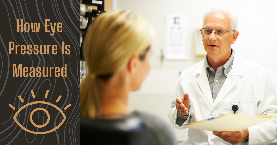
Patients are referring to air-puff or non-contact tonometry. Tonometry is the procedure used to measure eye pressure, and this is important for diagnosing and monitoring...
We commonly see patients who come in saying that their eyes are bleeding.
The patient is usually referring to the white part of their eye, which has turned bright red. The conjunctiva is the outermost layer of the eye and contains very fine blood...
