Blog
Visual Field
The visual field test is designed to see how well you see outside of the center part of your vision (peripheral vision).
When we test your vision on the basic eye chart it is only testing how well you see right in the center...
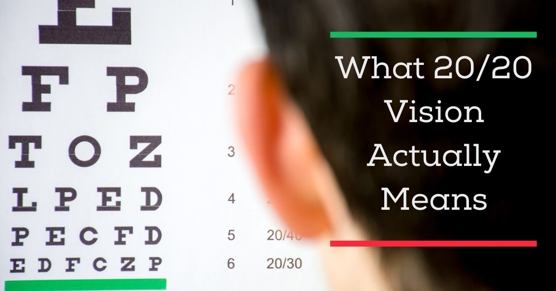
One of the most commonly asked questions in an eye exam comes right after the refraction, or glasses prescription check: “What is my vision?”
Almost invariably, people know the term “20/20”. In fact, it’s a measure of pride for many people....
