Blog

It's the summer and one of the most common questions eye doctors are asked is, “Is it safe to swim in my contact lenses?”
The answer we give is “NO!"
Do millions of people swim with their contact lenses? The answer is “Yes, they do, but it...
Read more: How to Ruin a Fun Day in the Water in One Easy Step
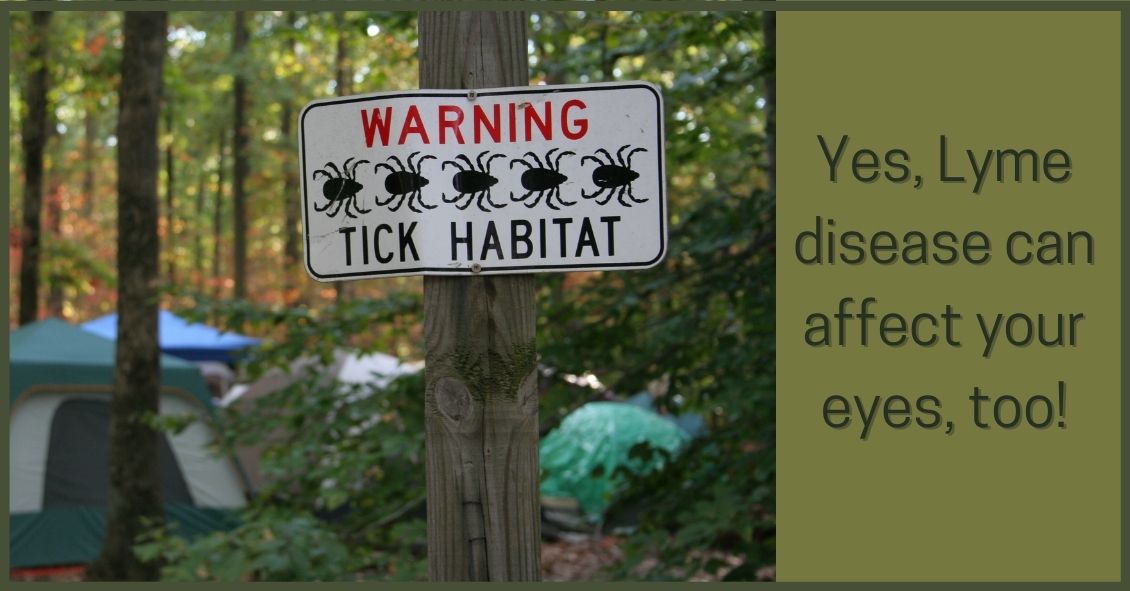
Lyme disease is an infection that is caused by a spirochete (a type of microorganism) called Borrelia burgdorferi. It is transmitted to humans by the bite of a deer tick.
The disease has a strong geographical incidence, being highly...
